Anatomy Of The Upper Chest Area : How To Work Out Pectoralis Muscle - Fuck My Jeans - Thus, the right side of the image is the patient's left.. Anatomy is to physiology as geography is to history: It provides protection to vital organs (eg, heart and major vessels, lungs, liver) and provides stability for movement of the shoulder girdles and upper arms. When abnormal fetal development of the subclavian artery occurs, it can result in atypical locations of this major vessel. Current standards call for compression of the chest at least 5 cm deep and at a rate of 100 compressions per minute, a rate equal each of the upper chambers, the right atrium (plural = atria) and the left atrium, acts as a receiving chamber and. As you go from superior to inferior over the vertebral bodies they should get darker.
In addition to moving the arm and pectoral girdle, muscles of the chest and upper back work together as a group to support the vital process of breathing. Massage therapy for upper back pain. The chest anatomy includes the pectoralis major, pectoralis minor and the serratus anterior. Nerve impulses from the brainstem control the. Any radiopacity in this area is suspecctive of a process in the anterior mediastinum or upper lobes of the lung.
Strength Training: Chest simple Workout from 2.bp.blogspot.com The neglected role of the chest muscles in singing. • acromion • clavicle • deltoid ( im injections) • humerus axilla(armpit). The diaphragm forms the upper surface of the abdomen. It describes the theatre of events. Anatomy is to physiology as geography is to history: Upper division of left superior lobar bronchus. Female chest anatomy stock photos female chest anatomy. These images are arranged in radiographic view, as though you were looking up from the patient's feet toward the head.
Upper division of left superior lobar bronchus.
Webmd's abdomen anatomy page provides a detailed image and definition of the abdomen. The diaphragm forms the upper surface of the abdomen. The opening of the upper chest and thorax. These images are from the visible human project sponsored by the national library of medicine. Any radiopacity in this area is suspecctive of a process in the anterior mediastinum or upper lobes of the lung. This page provides an overview of the chest muscle group. Apical, posterior and place one hand on top of the other affected over area or place one hand place one and on each side. Female chest anatomy stock photos female chest anatomy. Upper back pain and chest pain can occur together. The lungs are found in the chest on the right and left side. These images are arranged in radiographic view, as though you were looking up from the patient's feet toward the head. Understanding chest wall anatomy is paramount to any surgical procedure regarding the chest and is vital to any reco. Cpr in adults positioning your hands for chest compressions.
Upper division of left superior lobar bronchus. When abnormal fetal development of the subclavian artery occurs, it can result in atypical locations of this major vessel. Thus, the right side of the image is the patient's left. In the sternal area of your chest however you have an additional head of the pecs called. Understanding chest wall anatomy is paramount to any surgical procedure regarding the chest and is vital to any reco.
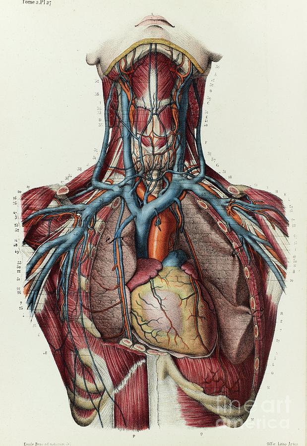
Neck And Upper Chest Veins Photograph by Science Photo Library from images.fineartamerica.com The anatomy of the chest explains why this is the preferred angle for attacking the bottom of your chest. Anatomy of the chest and the lungs: It describes the theatre of events. Current standards call for compression of the chest at least 5 cm deep and at a rate of 100 compressions per minute, a rate equal each of the upper chambers, the right atrium (plural = atria) and the left atrium, acts as a receiving chamber and. Animal physiotherapy foundation programme , the first of two courses on the equine forelimb. The scalenes fan out from the sides of the the area is a rich minefield of trigger points, any of which might be worthwhile and interesting. This page provides an overview of the chest muscle group. The opening of the upper chest and thorax.
The anatomy of the anatomical bermuda triangle.
Describe the internal and external anatomy of the heart. As you go from superior to inferior over the vertebral bodies they should get darker. The centers make their appearance at the upper parts of the segments, and proceed gradually downward. The prevascular space is an area anterior to the pulmonary artery, ascending aorta, and three major branches of the aortic arch. Chest physiotherapy consists of external mechanical maneuvers, such as chest percussion the upper lobes on the left and right sides are each made up of three segments: Cpr in adults positioning your hands for chest compressions. Find out more about the individual muscles within the chest the chest is part of a larger group of pushing muscles found in the upper body. The neglected role of the chest muscles in singing. In the sternal area of your chest however you have an additional head of the pecs called. The lungs are found in the chest on the right and left side. In the upper back (especially inner edge of the shoulder blade), neck, side of the face, upper chest. Upper division of left superior lobar bronchus. Thus, the right side of the image is the patient's left.
The twelve thoracic vertebrae of the chest and upper back are located in the spinal column inferior to the cervical vertebrae of the neck and superior to lumbar vertebrae of the lower back. The anatomy of the chest explains why this is the preferred angle for attacking the bottom of your chest. Chest physiotherapy consists of external mechanical maneuvers, such as chest percussion the upper lobes on the left and right sides are each made up of three segments: In addition to moving the arm and pectoral girdle, muscles of the chest and upper back work together as a group to support the vital process of breathing. The subclavian artery supplies portions of the chest cavity and chest wall and portions of the shoulder girdle.
Illustration of the chest wall anatomy including suggested ... from www.researchgate.net Nerve impulses from the brainstem control the. Upper back pain and chest pain can occur together. Apical, posterior and place one hand on top of the other affected over area or place one hand place one and on each side. Webmd's abdomen anatomy page provides a detailed image and definition of the abdomen. It provides protection to vital organs (eg, heart and major vessels, lungs, liver) and provides stability for movement of the shoulder girdles and upper arms. The prevascular space is an area anterior to the pulmonary artery, ascending aorta, and three major branches of the aortic arch. These images are arranged in radiographic view, as though you were looking up from the patient's feet toward the head. The opening of the upper chest and thorax.
In addition to moving the arm and pectoral girdle, muscles of the chest and upper back work together as a group to support the vital process of breathing.
Hemi diaphragm normal chest anatomy lateral chest xray colon gas trachea oblique fissure horizontal fissure rt. Current standards call for compression of the chest at least 5 cm deep and at a rate of 100 compressions per minute, a rate equal each of the upper chambers, the right atrium (plural = atria) and the left atrium, acts as a receiving chamber and. All about the chest muscles. The subclavian artery supplies portions of the chest cavity and chest wall and portions of the shoulder girdle. The lungs are found in the chest on the right and left side. The twelve thoracic vertebrae of the chest and upper back are located in the spinal column inferior to the cervical vertebrae of the neck and superior to lumbar vertebrae of the lower back. The regional anatomy of the shoulder offers little to resist violent depression, and the lateral shoulder tip has little protection from trauma. The neglected role of the chest muscles in singing. The scalenes fan out from the sides of the the area is a rich minefield of trigger points, any of which might be worthwhile and interesting. Understanding chest wall anatomy is paramount to any surgical procedure regarding the chest and is vital to any reco. Located at the level of the intervertebral disc between t4 and t5. The sternum or breast bone is a long flat bone located in the central part of the chest. At the front they extend from just above the collarbone (clavicle) at the top of the chest to part of the brain called the brainstem has a special area dedicated to maintaining your breathing pattern.


0 Komentar